An example of a pathology better understood thanks to brain imaging
Anatomical and functional abnormalities located in certain key regions of the brain for the establishment of social relationships (visual and auditory signals, language, face recognition) have been demonstrated in autistic children.
In positron emission tomography (PET), a significant bilateral decrease in cerebral blood flow at rest, located in the superior temporal sulcus (STS), was detected in nearly 80% of children with autism. The lower the cerebral blood flow, the more severe the autism (the higher the ADI score).
Correlation between DSC and severity of autism syndrome
Negative correlation with global ADI (p<0.001)
Anatomically, new methods of MRI analysis have confirmed the existence of structural abnormalities in these same superior temporal regions, including :
> a decrease in the area of the temporal sulcus with, again, a strong correlation between the area of the superior temporal sulcus and the severity of autistic symptoms.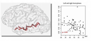
> a bilateral decrease in grey matter. In order to assess the specificity of this bitemporal abnormality, the results observed in children with autism were compared with those of healthy children and other subjects with Williams syndrome. Autism and Williams syndrome are two developmental disorders characterised by mirroring behaviours and symptoms. Social skills and communication are impaired in autism, whereas they are highly developed in Williams children, who are very sociable and have highly structured language. There is a gradient in grey matter density, which is lower in autistic children than in healthy subjects, who themselves have a lower density than children with Williams syndrome.
Brain activity was studied using functional MRI and a cortical area specialised in the recognition of the human voice, also located in the superior temporal sulcus, was objectified. Autistic patients do not activate this specific area for processing vocal information.
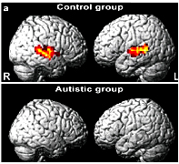
fMRI: absence of activation of the STS in response to voice
The superior temporal sulcus appears to play a central role. It is related to the brain regions involved in processing the sensory information necessary to analyse the dispositions and intentions of other individuals towards us (analysis of gaze or facial expression, recognition of the human voice, language) and making sense of the world around us. Abnormalities in the activation of all these regions have been observed in autistic subjects during tasks involving social interaction.
|
THE CONTRIBUTION OF THIS WORK IS MULTIPLE > diagnostic, raising the question of the interest of carrying out an MRI during the diagnostic and etiological assessment in autistic syndrome. > Therapeutic, with the prospect of developing specific re-education strategies that would aim, for example, to teach autistic children to decipher auditory and visual information, the pillars of their socialisation. But above all, a better understanding of the relationship between the disorders observed in autistic people and brain development will help to change society’s view of these patients and relieve the burden on families. |